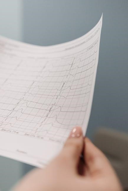
Rapid ECG Interpretation is a systematic guide by Dr. Dale Dubin, offering a clear, step-by-step approach to understanding electrocardiograms. It simplifies complex concepts for both novices and professionals, ensuring accurate and timely diagnoses in clinical settings.
1.1 Importance of ECG in Clinical Practice
The electrocardiogram (ECG) is a cornerstone in clinical practice, providing critical insights into cardiac function. It enables rapid identification of arrhythmias, ischemia, and life-threatening conditions, ensuring timely interventions. The ECG is essential for diagnosing acute myocardial infarction and monitoring treatment responses. Its ability to detect abnormalities, such as ST segment changes, makes it indispensable in emergency medicine. As a non-invasive tool, the ECG guides immediate patient care decisions, improving outcomes. Its significance extends to both primary and secondary care settings, making it a fundamental diagnostic instrument in modern healthcare.
1.2 Overview of Rapid ECG Interpretation
Rapid ECG Interpretation provides a systematic, step-by-step approach to analyzing electrocardiograms. Developed by Dr. Dale Dubin, this method emphasizes clarity and simplicity, breaking down complex concepts into digestible segments. The guide includes diagnostic criteria, instructive ECG examples, and practical protocols, ensuring accurate and efficient interpretation. It serves as a valuable resource for both novices and experienced professionals, offering a quick review for proficiency tests and board preparation. The third edition reflects updates in cardiology practice, making it a comprehensive yet concise tool for mastering ECG interpretation in clinical and emergency settings.
1.3 Historical Development of ECG Interpretation
The history of ECG interpretation began with Willem Einthoven’s invention of the electrocardiogram in 1903, revolutionizing cardiac diagnostics. Over the decades, advancements in technology and understanding of cardiac physiology refined ECG interpretation. Rapid ECG Interpretation by Dr; Dale Dubin builds on this foundation, offering a modern, systematic approach. The guide incorporates historical insights while addressing contemporary cardiology practices, making it a bridge between past discoveries and current clinical applications. This evolution ensures that ECG interpretation remains a vital, dynamic tool in healthcare, aiding accurate diagnoses and guiding timely interventions across generations of medical practice.
Basic Concepts of ECG
Understanding ECG basics involves recognizing key components like P, QRS, and T waves, and the ST segment. These elements help assess heart function and diagnose abnormalities efficiently in clinical settings.
2.1 Essential Components of an ECG
The essential components of an ECG include the P wave, QRS complex, T wave, and ST segment. The P wave represents atrial depolarization, while the QRS complex signifies ventricular depolarization. The T wave reflects ventricular repolarization, and the ST segment is the period between ventricular depolarization and repolarization. These elements are critical for assessing heart rhythm, rate, and potential abnormalities. Dr. Dale Dubin’s guide emphasizes the importance of these components in rapid and accurate ECG interpretation, providing a foundational understanding for both novices and experienced professionals to identify arrhythmias, ischemia, and other cardiac conditions effectively.
2.2 Understanding ECG Waves and Intervals
The ECG is composed of distinct waves and intervals that provide critical information about cardiac function. The P wave represents atrial depolarization, while the QRS complex signifies ventricular depolarization. The T wave reflects ventricular repolarization, and the ST segment is the flat line following the QRS complex. Key intervals include the PR interval (P wave to QRS onset), QT interval (QRS onset to T wave end), and QRS duration (ventricular depolarization time). Accurate measurement of these components is essential for identifying arrhythmias, bundle branch blocks, and ischemia, ensuring precise diagnoses in clinical practice.
2.3 The Significance of the Electrical Axis
The electrical axis represents the direction of the heart’s electrical activity, providing insights into ventricular depolarization. A normal axis ranges from -30° to +100°, while deviations may indicate conditions like left or right ventricular hypertrophy. Determined using limb leads (I, II, III, aVR, aVL, aVF), the axis helps identify structural heart diseases. An abnormal axis can signal fascicular blocks or hypertrophy, guiding further diagnostic steps. Understanding the axis is crucial for accurate ECG interpretation, as it reflects the heart’s electrical orientation and aids in detecting underlying cardiac abnormalities.

2.4 The Role of the ST Segment in Diagnosis
The ST segment is a critical component of the ECG, playing a vital role in diagnosing myocardial ischemia and infarction. ST segment elevation often indicates acute myocardial infarction, while depression may suggest ischemia. The ST segment’s deviation from the baseline helps identify these conditions early, enabling timely interventions. It is also useful in detecting other cardiac issues, such as pericarditis or ventricular hypertrophy. Accurate interpretation of the ST segment is essential for identifying life-threatening conditions and guiding appropriate treatment, making it a cornerstone in emergency and clinical cardiology settings.

Systematic Approach to ECG Interpretation
A step-by-step method ensures accurate ECG analysis, focusing on rate, rhythm, P waves, QRS complexes, and ST segments. This structured approach enhances diagnostic precision and clinical decision-making.
3.1 Step-by-Step Method for Accurate Interpretation
The step-by-step method in Rapid ECG Interpretation ensures a systematic approach to analyzing electrocardiograms. It begins with assessing the heart rate and rhythm, followed by examining P waves, QRS complexes, ST segments, and T waves. This structured process helps identify abnormalities such as arrhythmias, hypertrophy, or ischemia. By breaking down the ECG into its components, healthcare providers can accurately diagnose conditions like atrial fibrillation or myocardial infarction. This method is particularly useful for novices, as it builds confidence and proficiency in interpreting complex tracings. The step-by-step approach also serves as a refresher for experienced professionals, ensuring precise and timely patient care.
3.2 Identifying Rate and Rhythm

Identifying the heart rate and rhythm is the first step in ECG interpretation. The rate is calculated by measuring the time between consecutive R waves, with normal rates ranging from 60 to 100 beats per minute. Rhythm assessment involves determining if the heartbeat is regular or irregular. A regular rhythm has consistent R-R intervals, while an irregular rhythm, such as atrial fibrillation, lacks this consistency. Accurate identification of rate and rhythm is critical for diagnosing arrhythmias and guiding timely interventions. This step ensures a solid foundation for further analysis of P waves, QRS complexes, and other ECG components.
3.3 Analyzing P Wave Abnormalities
Analyzing P wave abnormalities is crucial for identifying atrial-related conditions. A normal P wave is upright in lead II, reflecting right atrial depolarization, with a duration of 0.08-0.11 seconds. Abnormalities include inverted or notched P waves, which may indicate left atrial enlargement or atrial fibrillation. In atrial fibrillation, P waves are absent, replaced by fibrillatory waves. Accurate P wave analysis helps diagnose conditions like atrial hypertrophy or arrhythmias, guiding appropriate interventions. This step in ECG interpretation requires careful measurement and correlation with clinical symptoms to ensure precise patient care and management.
3.4 Evaluating QRS Complex and Bundle Branch Blocks
The QRS complex represents ventricular depolarization, with a normal duration of ≤0.12 seconds. Abnormalities include widened QRS (>0.12s), indicating conduction delays. Bundle branch blocks (BBBs) occur when electrical impulses are delayed in the left (LBBB) or right (RBBB) bundle branches. LBBB shows a broad, notched R wave in lateral leads, while RBBB exhibits an rSR’ pattern in V1. These patterns are crucial for diagnosing conditions like ischemia or cardiomyopathy. Accurate evaluation of QRS and BBBs ensures timely identification of structural heart diseases, guiding appropriate interventions and improving patient outcomes in clinical practice.
3.5 Assessing ST Segment and T Wave Abnormalities
The ST segment and T wave are critical for diagnosing myocardial ischemia and infarction. ST segment elevation suggests acute MI, while depression indicates ischemia. T wave inversion may signify ischemia, ventricular hypertrophy, or electrolyte imbalances. The magnitude and direction of these changes are vital for accurate interpretation. Comparing these elements across leads helps localize abnormalities. Timely assessment of ST and T wave abnormalities is essential for identifying life-threatening conditions, enabling prompt interventions like reperfusion therapy. This step ensures precise diagnosis and guides urgent care, making it a cornerstone in rapid ECG interpretation and clinical decision-making.

Common ECG Abnormalities
Common ECG abnormalities include arrhythmias, ST segment changes, T wave inversion, and signs of hypertrophy or conduction disorders. These patterns are crucial for diagnosing cardiac conditions accurately and promptly.
4.1 Atrial and Ventricular Hypertrophy
Atrial and ventricular hypertrophy are key ECG findings that indicate enlarged heart chambers. Atrial enlargement is identified by tall P waves (>2.5mm) in leads II, III, and aVF, while ventricular hypertrophy is marked by increased QRS complex amplitude. Left ventricular hypertrophy (LVH) is the most common type, showing deep S waves in leads V1-V2 and tall R waves in leads V5-V6. The electrical axis may shift leftward in LVH. These changes are crucial for diagnosing conditions like hypertension or cardiomyopathy. Accurate interpretation of these patterns aids in identifying underlying cardiac disease and guiding further management.
4.2 Myocardial Infarction and Ischemia
Myocardial infarction (MI) and ischemia are critical conditions diagnosed via ECG. ST segment elevation indicates acute MI, while Q waves suggest prior infarction. Ischemia often presents as ST depression or T-wave inversion. These changes help identify affected coronary arteries and guide urgent interventions. Early ECG interpretation is vital for timely reperfusion therapy, improving patient outcomes. The electrocardiogram remains a cornerstone in detecting myocardial damage, enabling rapid diagnosis and treatment of life-threatening cardiac events.
4.3 Arrhythmias and Conduction Disorders
Arrhythmias and conduction disorders are commonly identified through ECG analysis. These conditions involve irregular heart rhythms or impaired electrical conduction. Atrial fibrillation, ventricular tachycardia, and heart blocks are key examples. The ECG helps differentiate between various arrhythmias by analyzing P-wave morphology, PR intervals, and QRS patterns. Conduction disorders, such as bundle branch blocks, are diagnosed by assessing QRS duration and axis deviation. Early detection of these abnormalities is critical for timely intervention, preventing complications like stroke or cardiac arrest. Rapid ECG interpretation is essential for identifying and managing these potentially life-threatening conditions effectively.
4.4 Miscellaneous Conditions Affecting ECG
Miscellaneous conditions such as electrolyte imbalances, hypothermia, and drug effects can significantly alter ECG readings. Hyperkalemia may cause tall, peaked T waves, while hypokalemia leads to flattened T waves and prolonged QT intervals. Hypothermia is associated with the Osborn wave, a distinctive J-shaped deflection. Digitalis effect manifests as scooped depressions in the ST segment. These changes are crucial for early diagnosis and treatment. Recognizing these patterns ensures accurate ECG interpretation, as they often indicate underlying systemic issues requiring immediate attention. Rapid identification of these abnormalities is vital for patient care and management.

Advanced Topics in ECG Interpretation
Advanced topics explore complex ECG concepts, including electrical axis deviations, fascicular blocks, and ECG changes in non-cardiac conditions like pulmonary embolism or electrolyte imbalances, enhancing diagnostic precision.
5.1 Electrical Axis and Fascicular Blocks
Electrical axis determination is crucial for identifying deviations that may indicate underlying cardiac conditions. A normal axis ranges from -30° to +100°, while left or right axis deviations suggest ventricular hypertrophy or bundle branch blocks. Fascicular blocks, such as left anterior or posterior fascicular block, are diagnosed by analyzing QRS complex patterns and ST segment shifts. These blocks often precede complete bundle branch blocks, emphasizing their importance in early detection. Understanding axis and fascicular block patterns enhances diagnostic accuracy, particularly in conditions like ischemia or cardiomyopathy, where timely intervention is critical. This systematic approach ensures precise identification of complex ECG abnormalities.
5.2 ECG Changes in Cardiac and Non-Cardiac Conditions
Electrocardiogram (ECG) changes are not limited to cardiac conditions; they also reflect systemic and non-cardiac disorders. Cardiac conditions like myocardial infarction, hypertrophy, and arrhythmias produce distinct ECG patterns. Non-cardiac conditions, such as electrolyte imbalances, pulmonary embolism, and hypothermia, also alter ECG traces. For instance, hyperkalemia causes tall, peaked T waves, while hypokalemia flattens T waves. Pulmonary embolism may show sinus tachycardia or right bundle branch blocks. Even neurological disorders, like subarachnoid hemorrhage, can induce ST-T wave changes. Recognizing these patterns is essential for accurate diagnosis and timely intervention, emphasizing the ECG’s role beyond cardiac care.
5.3 The Role of ECG in Emergency Medicine
ECG is a cornerstone in emergency medicine, enabling rapid identification of life-threatening conditions like arrhythmias, myocardial infarction, and ischemia. It provides immediate insights into cardiac function, guiding critical decisions such as thrombolytic therapy or defibrillation. In acute settings, ECG helps differentiate between cardiac and non-cardiac causes of symptoms like chest pain or syncope. Its quick interpretation is vital for triaging patients and initiating timely interventions. Educational resources like Rapid ECG Interpretation emphasize systematic approaches, ensuring emergency providers can accurately and swiftly interpret ECGs, improving patient outcomes in high-stakes situations.

Self-Assessment and Board Preparation
Rapid ECG Interpretation includes self-assessment quizzes and quick reference sheets, aiding healthcare professionals in exam preparation and proficiency tests. It ensures a systematic approach for accurate ECG analysis.
6.1 ECG Board Self-Assessment Quiz
Rapid ECG Interpretation features a comprehensive self-assessment quiz designed to test proficiency in ECG analysis. The quiz includes multiple-choice questions and diagnostic ECG tracings, simulating real exam conditions. It covers essential topics like arrhythmias, hypertrophy, and ischemia, helping users identify knowledge gaps. The quiz is complemented by quick reference sheets, enabling users to verify answers and refine their interpretation skills. This tool is invaluable for physicians and students preparing for board exams or proficiency tests, ensuring they can interpret ECGs accurately and confidently in clinical settings.
6.2 Tips for Proficiency Tests and Exams
Rapid ECG Interpretation provides practical tips for excelling in proficiency tests and exams. Emphasizing a systematic approach, it recommends practicing with sample ECGs and focusing on basic concepts. Users are advised to review diagnostic criteria and correlate ECG findings with clinical scenarios. Time management is stressed, with strategies for quickly identifying key abnormalities. The book also suggests using the included quick reference sheets for last-minute reviews. Regular practice with the self-assessment quiz is encouraged to build confidence and accuracy. These tips ensure healthcare professionals are well-prepared for exams and real-world applications.

Clinical Applications of Rapid ECG Interpretation
Rapid ECG Interpretation is a cornerstone in clinical practice, enabling quick identification of arrhythmias, ischemia, and life-threatening conditions. It ensures timely diagnostics and precise patient care in emergencies.
7.1 Timely Diagnostics and Interventions
Rapid ECG interpretation is crucial for identifying acute cardiac conditions, such as myocardial infarction and ischemia, enabling early intervention. It ensures timely diagnostics, improving patient outcomes by guiding immediate treatments like reperfusion therapy or arrhythmia management. This systematic approach streamlines decision-making, reducing delays in critical care settings. By providing clear, actionable insights, rapid ECG interpretation becomes a lifesaving tool, especially in emergencies where every minute counts. Its practical orientation ensures healthcare providers can act swiftly, enhancing the effectiveness of interventions and improving survival rates in high-stakes situations.
7.2 ECG in Primary and Secondary Care Settings
ECG interpretation plays a vital role in both primary and secondary care, serving as a first-line diagnostic tool for assessing cardiac conditions. In primary care, it aids in identifying patients with potential cardiac issues, enabling early referrals. In secondary care, it guides immediate interventions for acute conditions like myocardial infarction or arrhythmias. The systematic approach in Rapid ECG Interpretation ensures accuracy and efficiency, making it indispensable for healthcare providers. This tool bridges the gap between initial assessment and specialized care, enhancing patient outcomes through timely and informed decision-making.
Rapid ECG Interpretation has revolutionized cardiac diagnostics, offering a systematic approach to understanding electrocardiograms. Its evolution and future advancements promise enhanced accuracy and accessibility in healthcare settings.
8.1 The Evolution of ECG Interpretation
The evolution of ECG interpretation has transformed cardiac diagnostics, from manual readings to advanced digital analyses. Dr. Dale Dubin’s systematic approach in Rapid ECG Interpretation laid the groundwork for modern techniques, emphasizing step-by-step evaluation. Over the decades, the integration of AI and handheld devices has enhanced accuracy and accessibility. These advancements ensure that ECG interpretation remains a cornerstone in clinical practice, enabling timely and precise patient care across diverse healthcare settings.
8.2 Future Directions in ECG Technology
Future advancements in ECG technology aim to enhance accuracy, accessibility, and integration with modern healthcare systems. AI-driven algorithms are being developed to improve interpretation speed and precision, reducing human error. Handheld and wearable ECG devices are becoming more prevalent, enabling real-time monitoring and early detection of abnormalities. These innovations are expected to expand the role of ECGs in preventive care and remote patient management. Additionally, advancements in software and data analysis will further streamline the process, making rapid ECG interpretation more efficient and widely available for both clinicians and patients.