The cranial nerve examination is a fundamental skill in neurological assessment, enabling healthcare professionals to evaluate brain function and identify abnormalities․ This guide provides a structured approach, including PDF resources, to master the examination techniques for all twelve cranial nerves, ensuring accurate and efficient patient evaluation․
Overview of Cranial Nerves
The twelve pairs of cranial nerves originate from the brain and regulate essential functions, including movement, sensation, and involuntary actions․ They connect the brain to various structures, enabling control over activities like vision, hearing, and swallowing․ Each nerve has distinct roles, from transmitting sensory information to controlling motor functions․ Understanding their anatomy and functions is crucial for accurate neurological assessment and diagnosing lesions or abnormalities․
Importance in Neurological Assessment
The cranial nerve examination is vital for assessing brain function and identifying neurological deficits․ It helps diagnose conditions affecting the brainstem, nerves, and surrounding structures․ By evaluating sensory and motor functions, healthcare professionals can localize lesions and guide targeted treatments․ This skill is essential in clinical practice, particularly in OSCEs, where accurate assessment ensures proper patient care and management․
Preparation for the Exam
Proper preparation is key to a successful cranial nerve examination․ Begin by gathering essential equipment, such as a Snellen chart, ophthalmoscope, and reflex hammer․ Introduce yourself to the patient, explain the process, and ensure their comfort․ Position the patient appropriately, depending on the nerve being assessed․ Wash your hands and don PPE if necessary․ Organize your approach to systematically evaluate each cranial nerve, ensuring accuracy and efficiency in your assessment․

Cranial Nerve I: Olfactory Nerve
The olfactory nerve (CN I) is responsible for transmitting sensory information related to smell․ Assessment involves testing the patient’s ability to identify different odors, ensuring accurate cranial nerve examination․
Function and Assessment
The olfactory nerve (CN I) facilitates the sense of smell by transmitting sensory information from the nasal mucosa to the brain․ Assessment involves testing the patient’s ability to detect and identify different odors, such as coffee or vanilla, using each nostril separately․ This evaluation helps identify anosmia or hyposmia, which may indicate neurological or structural abnormalities affecting the cranial nerve pathways․
Common Abnormalities
Common abnormalities of the olfactory nerve include anosmia (loss of smell) or hyposmia (reduced smell), often due to trauma, infections, or neurological conditions․ Unilateral or bilateral impairment can indicate structural issues, such as nasal obstructions or brain tumors․ Early detection through smell testing is crucial for diagnosing underlying pathologies and guiding appropriate management strategies in clinical practice․
Cranial Nerve II: Optic Nerve
The optic nerve (CN II) is crucial for vision, transmitting visual information from the retina to the brain․ Its assessment involves testing visual acuity and visual fields, with ophthalmoscopy used to evaluate the optic disc for signs of atrophy or swelling, aiding in the detection of neurological conditions․
Visual Acuity and Field Testing
Assessing visual acuity involves using a Snellen chart to measure the sharpness of vision․ The patient reads letters with one eye covered at a time․ Visual field testing evaluates peripheral vision, often through confrontation, where the examiner compares their own fields to the patient’s․ Defects or blind spots may indicate optic nerve or brain pathway abnormalities, aiding in early detection of neurological conditions․
Ophthalmoscopy Examination
Ophthalmoscopy is a critical step in assessing the optic nerve (CN II)․ The examiner uses an ophthalmoscope to visualize the optic disc, retina, and macula․ Key observations include disc color, cup-to-disc ratio, and vessel caliber․ Abnormalities such as disc swelling (papilledema) or atrophy may indicate conditions like increased intracranial pressure or optic neuritis․ Proper positioning and patient cooperation are essential for a clear view and accurate assessment․
Cranial Nerve III: Oculomotor Nerve
The oculomotor nerve (CN III) controls eye movement, pupillary response, and eyelid elevation․ Examination includes assessing eye movements, pupillary reflexes, and checking for ptosis or diplopia․
Eye Movement and Pupillary Response
Assess eye movement by asking the patient to follow an object in all directions, testing the function of the medial rectus, inferior rectus, and inferior oblique muscles․ Evaluate pupillary response using the pupillary light reflex, noting constriction and reaction to light․ Abnormalities like ptosis or diplopia may indicate oculomotor nerve dysfunction, requiring further neurological evaluation․
Assessment Techniques
Assessment of the oculomotor nerve involves evaluating eye movements, pupillary reflexes, and lid elevation․ Use a penlight to test the pupillary light reflex, observing for constriction and reactivity․ Assess eye movement by having the patient track an object in all directions․ Note any nystagmus or limitations in movement․ Additionally, measure lid elevation and check for ptosis․ Abnormalities like anisocoria or impaired reflexes may indicate nerve dysfunction, guiding further neurological evaluation․

Cranial Nerve IV: Trochlear Nerve
The trochlear nerve (CN IV) controls the superior oblique muscle, essential for eye movement․ It is the thinnest and longest cranial nerve, with a unique intracranial course․
Function and Testing
The trochlear nerve (CN IV) controls the superior oblique muscle, enabling rotational eye movement․ Testing involves assessing eye movement, particularly looking medially and downward, to identify dysfunction or palsy, which may cause diplopia or impaired coordination․ Accurate examination ensures early detection of lesions affecting this nerve, guiding appropriate neurological interventions and patient care strategies effectively․
Common Lesions
The trochlear nerve (CN IV) is prone to lesions due to its thinness and long intracranial course․ Traumatic brain injury and microvascular ischemia are common causes․ Lesions often result in superior oblique muscle palsy, causing difficulty in downward and inward eye movement, leading to diplopia, especially when looking downward․ Patients may exhibit compensatory head tilting to alleviate symptoms․

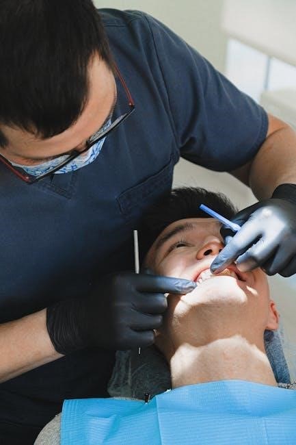
Cranial Nerve V: Trigeminal Nerve
The trigeminal nerve (CN V) manages sensory input and motor functions for the face․ It is divided into three branches: ophthalmic, maxillary, and mandibular․ Sensory functions include facial sensation, while motor functions control chewing muscles․ Examination involves assessing facial sensation, corneal reflex, and jaw movement to identify potential lesions or abnormalities affecting these functions․
Sensory and Motor Functions
The trigeminal nerve (CN V) has dual roles: sensory and motor․ Sensory functions include transmitting facial sensations like touch, pain, and temperature via its ophthalmic, maxillary, and mandibular branches․ The motor component controls the muscles of mastication, enabling chewing and jaw movements․ The corneal reflex, involving the ophthalmic and facial nerves, is also mediated by CN V, highlighting its critical role in both sensory perception and motor coordination of the face․
Examination Methods
Examination of the trigeminal nerve involves assessing both sensory and motor functions․ Sensory testing includes evaluating light touch, pain, and temperature sensation on the face using a cotton swab or a sharp object․ Motor function is assessed by examining the muscles of mastication, such as the temporalis and masseter muscles, during jaw clenching․ The corneal reflex, involving the ophthalmic branch and facial nerve, is also tested to ensure proper nerve function․

Cranial Nerve VI: Abducens Nerve
The abducens nerve controls lateral eye movement via the lateral rectus muscle․ Examination involves assessing eye abduction and checking for signs of dysfunction, such as medial strabismus or nystagmus․
The abducens nerve (CN VI) controls the lateral rectus muscle, enabling eye abduction․ Assessment involves testing lateral gaze by asking the patient to look sideways․ Observing for medial strabismus or nystagmus can indicate dysfunction․ A cranial nerve exam PDF guide provides detailed steps for evaluating CN VI, ensuring accurate identification of abnormalities and facilitating proper neurological assessment․
Abnormal Findings
Abnormal findings in CN VI assessment include medial strabismus, nystagmus, or inability to abduct the eye․ Lesions affecting CN VI may result from conditions like increased intracranial pressure, stroke, or tumors․ A cranial nerve exam PDF guide provides detailed criteria for identifying these abnormalities, ensuring accurate diagnosis and appropriate management of underlying neurological conditions․
Cranial Nerve VII: Facial Nerve
The facial nerve governs motor functions like facial expressions and sensory functions like taste․ A cranial nerve exam PDF provides detailed checklists for assessing these functions accurately․
Motor and Sensory Functions
The facial nerve controls motor functions like facial expressions and sensory functions, including taste for the anterior two-thirds of the tongue․ Damage can cause facial weakness or paralysis, affecting smile symmetry or eye closure․ Sensory impairment may reduce taste sensation․ A cranial nerve exam PDF provides structured checklists and detailed steps to assess these functions, ensuring accurate identification of abnormalities and guiding further diagnostic steps effectively․
Examination Techniques
Examination of the facial nerve involves assessing motor functions by observing facial symmetry and testing muscles of facial expression․ Patients are asked to smile, show teeth, or raise eyebrows․ Sensory function is evaluated by testing taste on the anterior tongue․ A cranial nerve exam PDF provides detailed steps and checklists to ensure thorough assessment, helping learners master these essential clinical skills effectively․
Cranial Nerve VIII: Vestibulocochlear Nerve
The vestibulocochlear nerve governs hearing and balance․ Assessment includes hearing tests with tuning forks and balance evaluation using the Romberg test․ A cranial nerve exam PDF outlines these methods․
Hearing and Balance Assessment
Assessing the vestibulocochlear nerve involves evaluating hearing and balance․ Hearing tests include pure tone audiometry and speech recognition․ Balance is assessed using the Romberg test and caloric reflex testing․ A cranial nerve exam PDF provides detailed steps for these evaluations, ensuring accurate identification of impairments related to vestibular or cochlear dysfunction․ These methods help diagnose conditions like unilateral hearing loss or vertigo․
Common abnormalities in cranial nerve exams include unilateral or bilateral palsies, such as facial weakness in cranial nerve VII lesions or impaired eye movement in cranial nerve III/IV/VI dysfunction․ Conditions like Bell’s palsy, acoustic neuromas, or multiple sclerosis can cause these deficits․ A cranial nerve exam PDF provides detailed checklists to identify and document these findings accurately during patient assessments․
Cranial Nerve IX: Glossopharyngeal Nerve
The glossopharyngeal nerve (CN IX) manages swallowing, taste, and salivation․ Testing involves assessing gag reflex, taste sensation, and swallowing ability․ A cranial nerve exam PDF provides detailed methods for evaluating these functions, ensuring accurate neurological assessments․
The glossopharyngeal nerve (CN IX) controls swallowing, taste sensation, and salivation․ Testing involves assessing the gag reflex, evaluating taste perception, and observing swallowing mechanics․ A cranial nerve exam PDF outlines these methods, ensuring thorough evaluation of CN IX’s functions; Abnormalities may indicate neurological issues, making precise testing crucial for accurate diagnosis and patient care․
Common lesions of the glossopharyngeal nerve (CN IX) often result from conditions like jugular foramen tumors or strokes․ These lesions can cause difficulty swallowing, loss of taste, and vocal cord weakness․ A cranial nerve exam PDF provides detailed checklists to identify such abnormalities, ensuring accurate diagnosis and appropriate management of CN IX-related pathologies․
Cranial Nerve X: Vagus Nerve
The vagus nerve (CN X) regulates vital functions like heart rate, digestion, and speech․ A cranial nerve exam PDF outlines its assessment, including vocal cord movement and gag reflex testing․
The vagus nerve (CN X) controls various involuntary functions, including heart rate, digestion, and respiratory rate․ Assessment involves evaluating speech, gag reflex, and vocal cord movement․ A cranial nerve exam PDF provides detailed steps for testing these functions, ensuring accurate identification of abnormalities․ Proper technique is essential to avoid missing potential lesions or impairments in this critical nerve․
Abnormal findings in the vagus nerve examination may include hoarseness, difficulty swallowing, or an absent gag reflex․ These signs can indicate nerve damage or lesions․ A cranial nerve exam PDF highlights key abnormalities, such as vocal cord paralysis or altered speech, aiding in early detection of conditions like stroke or neuropathy․ Recognizing these findings is crucial for accurate diagnosis and timely intervention․
Cranial Nerve XI: Spinal Accessory Nerve
The spinal accessory nerve controls neck movements and sternocleidomastoid muscle function․ Assessment involves testing strength and range of motion, with abnormalities indicating potential nerve damage․ A cranial nerve exam PDF provides detailed evaluation techniques for accurate diagnosis․
The spinal accessory nerve governs the sternocleidomastoid and trapezius muscles, essential for neck rotation and shoulder movement․ Testing involves assessing muscle strength and range of motion․ Abnormalities, such as weakness or reduced mobility, may indicate nerve damage․ A cranial nerve exam PDF outlines detailed methods for evaluating these functions, ensuring accurate identification of potential lesions or impairments during neurological assessments․
Common abnormalities include unilateral or bilateral palsies, such as weakness or paralysis of specific cranial nerve-innervated muscles․ Lesions affecting multiple nerves may indicate conditions like cerebellopontine angle tumors or jugular foramen syndrome․ Bilateral palsies of nerves IX, X, and XI often suggest bulbar or pseudobulbar palsy․ A cranial nerve exam PDF provides detailed insights into recognizing these patterns and their clinical implications for accurate diagnosis․
Cranial Nerve XII: Hypoglossal Nerve
The hypoglossal nerve controls tongue movements․ Assessment involves checking for wasting, fasciculations, and deviation when the tongue is protruded, as detailed in the cranial nerve exam PDF․
The hypoglossal nerve (CN XII) controls tongue movements, including protrusion, lateral deviation, and speech articulation․ Assessment involves observing tongue protrusion for deviation, testing strength against resistance, and checking for wasting or fasciculations․ A detailed cranial nerve exam PDF guide outlines these steps, ensuring accurate evaluation of motor function and early detection of impairments like atrophy or weakness․
Common lesions of the hypoglossal nerve (CN XII) include unilateral palsies, often due to trauma, tumors, or stroke․ These lesions can cause tongue weakness, wasting, or deviation․ Conditions like amyotrophic lateral sclerosis (ALS) may also affect CN XII, leading to progressive muscle atrophy․ A detailed cranial nerve exam PDF guide helps identify these lesions, emphasizing clinical signs and their implications for diagnosis and management․
Common Cranial Nerve Lesions
Common cranial nerve lesions include tumors, strokes, and traumatic injuries․ These often affect multiple nerves, such as cerebellopontine angle tumors impacting CN V, VII, and VIII․ A cranial nerve exam PDF provides detailed insights into identifying and managing these conditions effectively․
Multiple Cranial Nerve Lesions
Multiple cranial nerve lesions often result from conditions like tumors, strokes, or infections․ For instance, cerebellopontine angle tumors can affect CN V, VII, and VIII, while jugular foramen lesions impact CN IX, X, and XI․ A cranial nerve exam PDF provides a systematic approach to identifying these patterns, aiding in precise diagnosis and management of complex neurological cases․
Unilateral and Bilateral Palsies
Unilateral palsies of CN V, VII, and VIII suggest cerebellopontine angle lesions, while CN IX, X, and XI palsies indicate jugular foramen involvement․ Bilateral palsies of CN X, XI, and XII may signify bulbar palsy (LMN changes) or pseudobulbar palsy (UMN signs)․ A cranial nerve exam PDF helps differentiate these conditions, guiding accurate diagnosis and management of complex neurological disorders․

OSCE Guide to Cranial Nerve Examination
This cranial nerve examination guide provides a clear, step-by-step approach for OSCE preparation, including a video demonstration and detailed techniques to assess all twelve cranial nerves effectively․
Step-by-Step Approach
A structured cranial nerve examination begins with introducing yourself, washing hands, and positioning the patient․ Assess each cranial nerve systematically, starting with CN I (olfactory) and proceeding to CN XII (hypoglossal)․ Use tools like a Snellen chart for visual acuity and a tongue depressor for motor function․ Document findings thoroughly and correlate with patient symptoms for accurate diagnosis․ This method ensures a comprehensive and efficient evaluation․
Video Demonstration
Video demonstrations provide a visual guide to mastering the cranial nerve examination, offering step-by-step instruction for each nerve assessment․ These resources complement written guides, making complex techniques easier to understand․ Ideal for medical students and professionals, videos often include practical examples and tips for real-world application, enhancing learning and clinical proficiency in neurological assessment․
Resources and Downloads
Access comprehensive cranial nerve exam PDF guides and checklists, offering detailed charts, exercises, and step-by-step instructions․ These resources are ideal for medical professionals and students seeking structured learning tools․
Cranial Nerve Exam PDF Guides
Cranial nerve exam PDF guides provide comprehensive resources for mastering neurological assessments․ These guides include detailed charts, exercises, and step-by-step instructions, making them ideal for medical students and professionals․ They offer systematic approaches to evaluating all twelve cranial nerves, ensuring accurate and efficient patient evaluations․ Downloadable checklists and visual aids enhance learning and clinical application, making these guides indispensable tools for healthcare education and practice․
Comprehensive Checklists
Comprehensive checklists for cranial nerve exams are essential tools for healthcare professionals and students․ These detailed lists cover all twelve cranial nerves, ensuring no step is missed during assessment․ They include mnemonics, diagrams, and step-by-step instructions to aid memory and improve accuracy․ By following these checklists, clinicians can systematically evaluate sensory, motor, and reflex functions, enhancing diagnostic precision and patient care outcomes in neurological examinations․
Mastery of the cranial nerve examination is essential for accurate neurological assessment․ Utilizing resources like cranial nerve exam PDF guides ensures a systematic approach, enhancing clinical skills․ These tools provide clear instructions and checklists, aiding in identifying abnormalities and improving patient outcomes․ By following these guidelines, healthcare professionals can confidently perform thorough examinations, ultimately advancing diagnostic precision and patient care in neurological practice․